

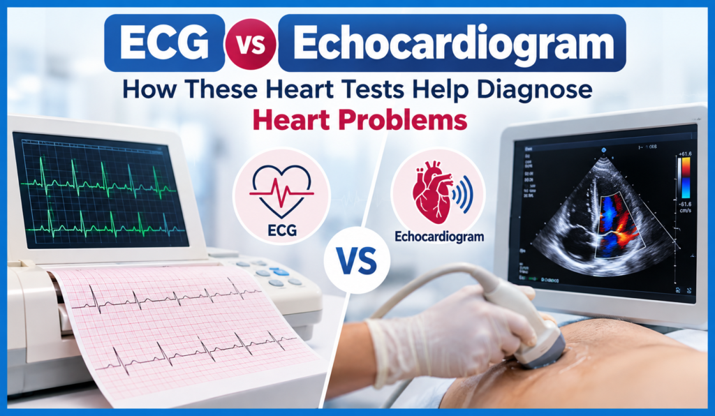
ECG vs Echocardiogram – Heart tests play an important role in detecting, and evaluating different heart conditions. There are two commonly recommended tests. One is ECG, and another one is echocardiogram. Although both are used to assess heart health, but they provide different types of information. An ECG records the heart’s electrical activity, while an echocardiogram uses ultrasound to create images of the heart’s structure, and movement.
This article briefly explains and helps in understanding the difference between these tests. This helps the patients know why their doctor may recommend one, or both.
ECG
An electrocardiogram is a quick, non-invasive test that records the electrical signals produced by the heart. These signals control the heartbeat, and help the heart contract in a coordinated way. During an ECG, small electrodes are placed on the chest, arms, and legs. These electrodes detect the heart’s electrical activity, and display it as a waveform on a monitor, or printout. An ECG usually takes only a few minutes, and does not require needles, or anaesthesia.
ECG identifies
- Abnormal heart rhythms
- Fast, or slow heart rates
- Certain signs of reduced blood flow to the heart
- Previous, or ongoing heart damage
- Electrical conduction problems
- Abnormalities related to the heart’s chambers
Echocardiogram or Echo
An echo is an ultrasound examination of the heart. It uses high frequency sound waves to produce moving images of the heart. It provides the movement of heart and its structure in image format which ECG cannot.
An echocardiogram can allow a doctor to evaluate the following
- Heart chambers
- Heart valves
- Heart muscle movement
- Pumping function
- Blood flow through the heart
- The size, and structure of the heart
- Certain abnormalities around the heart
ECG vs Echocardiogram - What is the difference?
ECG – Focuses primarily on the heart’s electrical activity, and rhythm
Echocardiogram – Focuses primarily on the heart’s structure, movement, pumping function, and blood flow
In simple terms, an ECG helps the doctors, how the heart’s electrical system is working, while an echocardiogram shows how the heart is built, and how it is functioning mechanically.
When ECG is recommended?
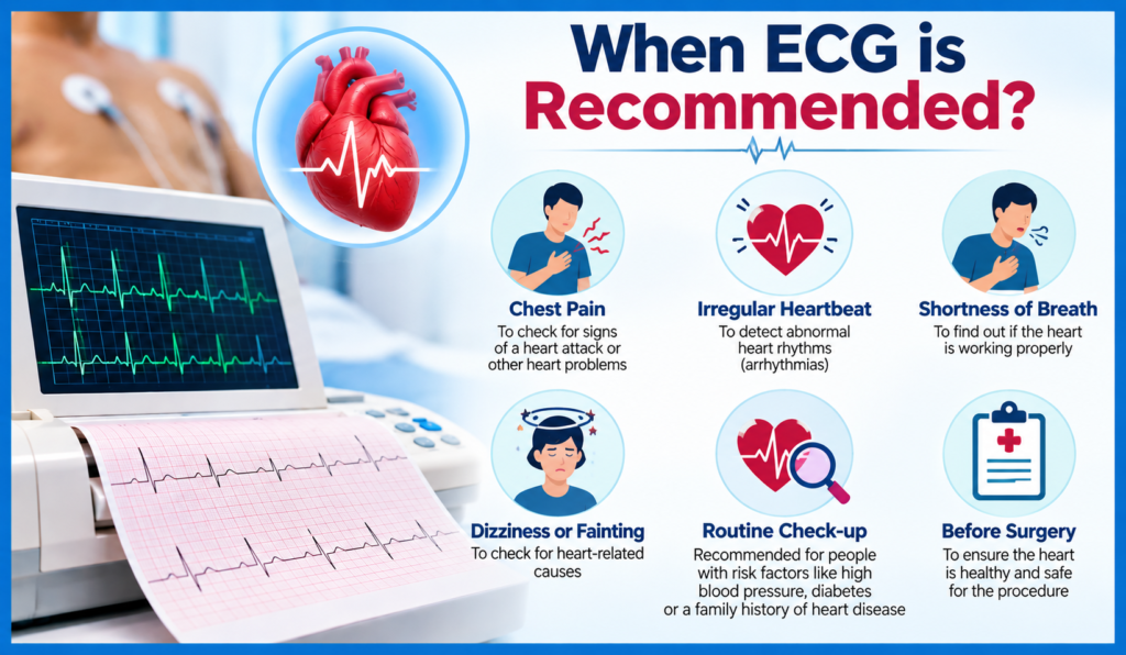
Doctors may recommend an ECG, when a patient has symptoms that could be related to abnormal heart activity. An ECG may also be performed as part of a general cardiac evaluation, or before certain procedures, depending on the patient’s circumstances.
Common reasons include
- Chest discomfort
- Palpitations, or a feeling of a racing heartbeat
- Dizziness
- Fainting
- Shortness of breath
- Unexplained fatigue
- Irregular pulse
- Monitoring certain known heart conditions
When is an Echocardiogram recommended?
An echocardiogram may be recommended when a doctor needs more detailed information about the heart’s structure, and function. An echo may also be used to monitor an existing heart condition, and assess how the heart responds to the treatment.
Echo helps evaluate the following
- Heart valve problems
- Heart muscle disorders
- Reduced pumping function
- Structural abnormalities
- Certain congenital heart conditions
- Fluid around the heart
- Some causes of breathlessness, or swelling
How Is an ECG performed?
An ECG is a simple, and painless procedure.
The process usually involves
- Preparation
The patient may be asked to lie comfortably, and remain still. - Electrode Placement
Small adhesive electrodes are placed on specific areas of the chest, and limbs. - Recording
The ECG machine records the heart’s electrical signals for a short period. - Completion
The electrodes are removed after the recording is complete. The patient can generally return to normal activities immediately unless another procedure is planned.
How is an Echocardiogram performed?
- Preparation
A standard echocardiogram is usually performed while the patient lies on an examination bed. - Application of Gel
The healthcare professional applies ultrasound gel to the chest, and moves a device called a transducer over different areas of the chest. - Recording
The device sends sound waves into the body, and receives echoes that are converted into images. The images can show the heart beating in real time. - Completion
Depending on the type of echocardiogram, additional preparation may sometimes be required.
Are these tests painful?
Both ECG, and standard echocardiography are generally non-invasive.
An ECG may cause minor discomfort when adhesive electrodes are removed from the skin. An echocardiogram may involve mild pressure from the ultrasound transducer against the chest, but it is generally not painful.
Specialized forms of echocardiography may involve additional procedures, so patients should discuss preparation, and potential discomfort with their healthcare professional.
Both ECG, and Echocardiogram be done together?
Yes. In some situations, doctors may recommend both tests. Because they provide complementary information.
For example, a patient experiencing palpitations may have an ECG to assess heart rhythm, while an echocardiogram may be used to evaluate the heart’s structure, and pumping function.
Using both tests can give doctors a broader understanding of certain heart problems.
Whether ECG, or Echocardiogram is better?
There is no single test that is better for every situation. ECG, and echocardiogram serve different purposes.
An ECG is particularly useful for assessing electrical activity, and rhythm, whereas an echocardiogram is useful for evaluating heart structure, movement, valves, and pumping function.
The appropriate test depends on the patient’s symptoms, medical history, physical examination, and what the doctor needs to investigate.
What happens after the tests?
After an ECG, or echocardiogram, a healthcare professional reviews the results along with the patient’s symptoms, and other clinical information.
An abnormal result does not necessarily mean that a serious heart condition is present. Further evaluation may sometimes be needed to understand the finding.
Depending on the situation, a doctor may recommend additional tests such as blood tests, stress testing, Holter monitoring, or other cardiac investigations.
When should you see a doctor?
Do not ignore persistent, or unexplained heart-related symptoms. Sudden severe chest pain, severe breathing difficulty, fainting, or other signs of a possible heart emergency require immediate medical attention.
Medical evaluation is particularly important if you experience
- New, or severe chest pain
- Difficulty breathing
- Fainting, or near-fainting
- Persistent palpitations
- Unusual swelling
- Severe, or unexplained fatigue
- A newly noticed irregular heartbeat
FAQs about ECG vs Echocardiogram
- What is the main difference between ECG, and echocardiogram?
An ECG records the heart’s electrical activity, while an echocardiogram uses ultrasound to examine the heart’s structure, and function. - Is an ECG painful?
No. An ECG is generally painless, and usually takes only a few minutes. - Does an echocardiogram show blocked arteries?
A standard echocardiogram does not directly show all coronary artery blockages. Other tests may be needed to evaluate suspected coronary artery disease. - Can I have both an ECG, and echocardiogram?
Yes. Doctors may recommend both because they provide different, and complementary information about the heart. - Which test should I take for heart problems?
The appropriate test depends on your symptoms, medical history, and clinical examination. A doctor can recommend the most suitable test.